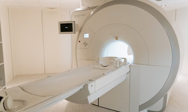
In today’s world of medicine, getting the proper diagnosis quickly is important. One tool that doctors use a lot for this is called a Computed Tomography (CT) scan. These scans have changed how people figure out what’s wrong with people. They give detailed pictures that help people plan the best treatment. This article discusses how CT scans work and why they’re so helpful in modern healthcare.
Understanding It
A CT scan is a special way of taking pictures of the body. It blends X-ray technology with computers to make detailed pictures of body sections. Unlike regular X-rays that show things in two dimensions, CT scans generate 3D images, helping doctors see the inside from different angles. This helps them understand how different parts fit together, which is essential for finding out what’s wrong and how to treat it.
Principle of Operation
CT scans use X-rays that get absorbed differently by different body parts. Solid things such as bones absorb a lot, so they look white in the pictures. Soft things, such as muscles and organs, take in less, so they look darker. To make the pictures, a machine with a spinning X-ray takes lots of photos from different sides of your body. A computer then puts all these pictures together to show slices of what’s inside you.
Diagnostic Precision
As various renowned sources, including the Inside Radiology website, will tell you, CT scans have this incredible superpower: they can create super detailed pictures of what’s inside your body. It’s as if having a superhero X-ray vision but for doctors. These pictures show everything so clearly that doctors can spot things such as broken bones, lumps that shouldn’t be there, messed up blood vessels, and even when your organs are acting all wonky. So yeah, CT scans are the ultimate medical detectives!
Clinical Applications
CT scans find applications across various medical specialties due to their versatility and diagnostic accuracy. In emergency medicine, CT scans are often employed to rapidly assess trauma patients for internal injuries and bleeding. In oncology, these scans aid in tumour staging and monitoring treatment response. CT angiography visualizes blood vessels and identifies potential blockages or aneurysms, helping prevent cardiovascular events.
Contrast-Enhanced CT Scans
To enhance the visibility of specific structures and abnormalities, contrast agents may be administered to the patient before the CT scan. These contrast agents contain compounds that absorb X-rays differently from surrounding tissues, highlighting specific areas of interest. Contrast-enhanced methods are commonly used in imaging blood vessels, the brain, and the abdomen. However, it’s essential to note that some patients may be allergic to these contrast agents, necessitating careful consideration before administration.
Reducing Radiation Exposure
While CT scans provide invaluable diagnostic information, there is a concern regarding the potential radiation exposure to patients. X-rays, which are ionizing radiation, can have cumulative harmful effects on cells if not controlled. To mitigate this risk, modern CT scanners are equipped with dose-reduction techniques. These techniques include automatic exposure control, which adjusts the radiation dose based on the patient’s size and the area being imaged, and iterative reconstruction algorithms that enhance image quality while reducing radiation dose.
The world of medical testing is constantly changing, and CT scans are the rock-solid foundation that keeps everything together. These scans are the magic windows into human bodies, showing all the nitty-gritty details. Doctors rely on them because they can make 3D pictures that help determine what’s going wrong. It’s as if having a super detective tool that spots various health issues.
